


ņä£ ļĪĀ
ĒÖöņāØļ░® ņāüĒÖ® ļ░Å Ēģīļ¤¼ ļ¬®ņĀüņ£╝ļĪ£ ņé¼ņÜ®ļÉśļŖö ĒÖöĒĢÖņ×æņÜ®ņĀ£ņØś ņŻ╝ņÜö ņØĖņ▓┤ ņ£Āņ×ģ Ļ▓ĮļĪ£ļŖö ĒśĖĒØĪ ļ░Å Ēö╝ļČĆĻ░Ć ļÉĀ ņłś ņ׳ļŗż. ĻĘĖ ņżæņŚÉņä£ ņŗĀĻ▓Įņ×æņÜ®ņĀ£(nerve agents)ņÖĆ ņłśĒżņ×æņÜ®ņĀ£(vesicating agent)ņØś Ļ▓ĮņÜ░, ņĢĪņ▓┤ ņāüĒā£ļĪ£ Ēö╝ļČĆ Ēæ£ļ®┤ņŚÉ ņśżņŚ╝ļÉĀ Ļ▓ĮņÜ░ ĻĘ╣ņåīļ¤ēņŚÉ ņØśĒĢ┤ņä£ļÅä ņé¼ļ¦ØņŚÉ ņØ┤ļź╝ ņłś ņ׳ļŖöļŹ░, ņŗĀĻ▓Įņ×æņÜ®ņĀ£ VXņØś Ļ▓ĮņÜ░ Ēö╝ļČĆņŚÉ ņśżņŚ╝ ņŗ£ ņ▓┤ņżæ 70 kg ņä▒ņØĖņØä ĻĖ░ņżĆņ£╝ļĪ£ ņżæĻ░ä ņ╣śņé¼ļ¤ē(LD50)ņØĆ ņĢĮ 10 mgņŚÉ ļČłĻ│╝ĒĢśļŗż(0.14 mg/kg)[1]. ļö░ļØ╝ņä£ ĒÖöĒĢÖņ×æņÜ®ņĀ£ņØś Ēö╝ļČĆ ņ╣©Ēł¼ļź╝ ņ¢ĄņĀ£ĒĢĀ ņłś ņ׳ļŖö Ēö╝ļČĆļ│┤ĒśĖņĀ£ ņŚ░ĻĄ¼Ļ░£ļ░£ņØä ņ£äĒĢśņŚ¼ ņ¦ĆĻĖłĻ╣īņ¦Ć ļ¦ÄņØĆ ņŚ░ĻĄ¼Ļ░Ć ņ¦äĒ¢ēļÉśņ¢┤ ņÖöļŗż[2-4]. ĻĘĖļ¤¼ļ»ĆļĪ£ ņØ┤ļ¤¼ĒĢ£ Ēö╝ļČĆļ│┤ĒśĖņĀ£ņØś ņŚ░ĻĄ¼Ļ░£ļ░£ņØä ņ£äĒĢśņŚ¼ ņ×æņÜ®ņĀ£ņØś Ēö╝ļČĆĒł¼Ļ│╝ļÅä ņĖĪņĀĢ ļ░Å Ēö╝ļČĆļ│┤ĒśĖņĀ£ņØś ņĀĢļ¤ēņĀüņØĖ ņä▒ļŖźĒÅēĻ░ĆļŖö ļ¦żņÜ░ ņżæņÜöĒĢśļŗż. ņØ┤ļź╝ ņ£äĒĢśņŚ¼ ļ»ĖĻĄŁ USAMRICD(US Army Medical Research Institute of Chemical Defense)ņŚÉņä£ļŖö M8 ĒÖöĒĢÖņ×æņÜ®ņĀ£ ĒāÉņ¦Ćņ¦Ć(detection paper)ļź╝ ņé¼ņÜ®ĒĢśņśĆļŖöļŹ░,[5] M8ĒāÉņ¦Ćņ¦ĆļŖö ņĢĪņ▓┤ņä▒ ņŗĀĻ▓Į ļ░Å ņłśĒżņ×æņÜ®ņĀ£ņÖĆ ņĀæņ┤ēļÉśļ®┤ ņāēņāüņØ┤ ļ│ĆĒĢśņŚ¼ ņ£ĪņĢłņ£╝ļĪ£ ņēĮĻ▓ī ņŗØļ│äĒĢĀ ņłś ņ׳ļŖö ņøÉļ”¼ļź╝ ņØ┤ņÜ®ĒĢśņŚ¼, ĒāÉņ¦Ćņ¦Ć Ēæ£ļ®┤ ņ£äņŚÉ Ēö╝ļČĆļ│┤ĒśĖņĀ£ļź╝ ļ░öļź┤Ļ│Ā ĻĘĖ ņ£äņŚÉ ņ×æņÜ®ņĀ£ļź╝ Ļ░ĆĒĢśļ®┤ ņ×æņÜ®ņĀ£Ļ░Ć ļ│┤ĒśĖņĀ£ ņĖĄņØä Ēł¼Ļ│╝ ņŗ£ ĒāÉņ¦Ćņ¦ĆņØś ņāēņāüļ│ĆĒÖöĻ░Ć ņŗ£ņ×æļÉśļŖö ņŗ£Ļ░ä(onset time)ņØä ņĖĪņĀĢĒĢ©ņ£╝ļĪ£ņä£ ļ│┤ĒśĖņĀ£ņØś ņä▒ļŖźņØä ņŖżĒü¼ļ”¼ļŗØĒĢśņśĆļŗż[5]. ļ│Ė ņŚ░ĻĄ¼ĒīĆņØĆ M8ĒāÉņ¦Ćņ¦Ć ļ│┤ļŗż ļ░śņØæņŗ£Ļ░ä(response time)ņØ┤ ļŹöņÜ▒ ļ╣ĀļźĖ[6] M9ĒāÉņ¦Ćņ¦Ćļź╝ ņé¼ņÜ®ĒĢśņŚ¼ ļ¦ÄņØĆ Ēøäļ│┤ļ¼╝ņ¦ł(ņĪ░ņä▒ļ¼╝)ņŚÉ ļīĆĒĢ£ ņŖżĒü¼ļ”¼ļŗØ ņŗ£ĒŚśņØä ņŗżņŗ£ĒĢ£ ļ░öĻ░Ć ņ׳ļŗż[7]. ĻĘĖļ¤¼ļéś ņŗżņĀ£ļĪ£ ĒÖöĒĢÖņ×æņÜ®ņĀ£Ļ░Ć ņāØņ▓┤ Ēö╝ļČĆņŚÉ ņśżņŚ╝ļÉśņŚłņØä Ļ▓ĮņÜ░ņŚÉ Ēö╝ļČĆĒł¼Ļ│╝ļÅäļź╝ ņĖĪņĀĢĒĢśĻĖ░ ņ£äĒĢśņŚ¼ ņŗĀĻ▓Įņ×æņÜ®ņĀ£ņØś Ļ▓ĮņÜ░ļŖö ĒśłņĢĪ ņżæņØś ĒÜ©ņåī cholinesteraseņØś ĒÖ£ņä▒ļÅäļź╝ ņĖĪņĀĢ(in-vivo test)ĒĢ©ņ£╝ļĪ£ņä£ ņ×æņÜ®ņĀ£ņØś Ēö╝ļČĆĒł¼Ļ│╝ļÅäļź╝ ņĀĢļ¤ēņĀüņ£╝ļĪ£ ņĖĪņĀĢņØ┤ Ļ░ĆļŖźĒĢśļŗż[8]. ĻĘĖļ¤¼ļéś ņŗżĒŚśļÅÖļ¼╝ņØś ņāØļ”¼ņāüĒā£ļéś Ļ░£ņ▓┤ ņ░©ņØ┤ņŚÉ ņØśĒĢ£ ņśüĒ¢ź ņÜöņØĖņØ┤ ļ¦Äņ£╝ļ»ĆļĪ£ ņĀĢļ░ĆĒĢśĻ▓ī Ēö╝ļČĆĒł¼Ļ│╝ļÅäļź╝ ņĖĪņĀĢĒĢśĻĖ░ņŚÉļŖö ĒĢ£Ļ│äĻ░Ć ņ׳ļŗż. ļśÉĒĢ£ ļŗżļźĖ ĒÖöĒĢÖņ×æņÜ®ņĀ£ņØś Ļ▓ĮņÜ░ļÅä ņŗżĒŚśļÅÖļ¼╝ Ēö╝ļČĆņŚÉ ņ×æņÜ®ņĀ£ļź╝ ņśżņŚ╝ņŗ£ĒéżĻ│Ā Ļ▓Įņŗ£ļ│äļĪ£ ĒśłņĢĪņØä ņ▒äņĘ©ĒĢśņŚ¼ Ēö╝ļČĆļź╝ Ēł¼Ļ│╝ĒĢ£ ņ×æņÜ®ņĀ£ņØś ļåŹļÅäļź╝ ņĀĢļ¤ēļČäņäØĒĢĀ ņłśļŖö ņ׳ņ¦Ćļ¦ī ņāØņ▓┤ ļé┤ņŚÉ ņĪ┤ņ×¼ĒĢśļŖö Ļ░üņóģ ļČäĒĢ┤ĒÜ©ņåīņŚÉ ņØśĒĢ┤ Ļ░ÉņåīļÉśļŖö ņ×æņÜ®ņĀ£ņØś ņ¢æņØä ņĀĢĒÖĢĒ׳ ņĢīĻĖ░ ņ¢┤ļĀĄļŗżļŖö ļŗ©ņĀÉņØ┤ ņ׳ļŗż. ļö░ļØ╝ņä£ ņśżļל ņĀäļČĆĒä░ ņŗżĒŚśļÅÖļ¼╝ņØś Ēö╝ļČĆņĪ░ņ¦ü ņĀłĒÄĖņØä ņé¼ņÜ®ĒĢśņŚ¼ in-vitroņŚÉņä£ ņĢĮļ¼╝ņØś Ēö╝ļČĆ Ēł¼Ļ│╝ļÅäļź╝ ņĀĢļ¤ēņĀüņ£╝ļĪ£ ĒÅēĻ░ĆĒĢśļŖö ļ░®ļ▓ĢņØ┤ ņŗ£ļÅäļÉśņŚłļŗż[9]. ĻĘĖ Ēøä ņØ┤ ļ░®ļ▓ĢņØś Ēæ£ņżĆĒÖöļź╝ ņ£äĒĢśņŚ¼ OECD Ļ░ĆņØ┤ļō£ļØ╝ņØĖ[10]ņØ┤ ļéśņÖöĻ│Ā ņłśļ¦ÄņØĆ ņŚ░ĻĄ¼ņ×ÉļōżņØ┤ ņØ┤ Ļ░ĆņØ┤ļō£ļØ╝ņØĖņØä ĒåĀļīĆļĪ£ Ļ▓ĮĒö╝ņä▒ ņ¦ĆņåŹņä▒ ņĢĮļ¼╝(TDDS: Transdermal Drug Delivery System), ĒŖ╣Ē׳ Ēö╝ļČĆļČĆņ░®ĒśĢ Ēī©ņ╣śņØś ņĢĮļ¼╝ ĒØĪņłśņŚÉ ļīĆĒĢ£ ņĢĮļ¼╝ļÅÖļĀźĒĢÖņĀü(pharmaco-dynamics)ņØĖ ņŚ░ĻĄ¼ļź╝ ņłśĒ¢ēĒĢśņśĆļŗż[11-13]. ņØ┤ ņŗżĒŚśļ░®ļ▓ĢņØĆ ņØĖņ▓┤ņŚÉ ņśüĒ¢źņØä ņżä ņłś ņ׳ļŖö Ļ░üņóģ ņ£äĒĢ┤ļ¼╝ņ¦łņØ┤ ņØĖņ▓┤ Ēö╝ļČĆļź╝ ņ¢╝ļ¦łļéś ĒåĄĻ│╝ĒĢĀ ņłś ņ׳ļŖöĻ░Ćļź╝ ļŗ©ņ£äņŗ£Ļ░äļ│äļĪ£ ņĀĢļ¤ēņĀüņ£╝ļĪ£ ĒÅēĻ░ĆĒĢĀ ņłś ņ׳ļŖö in-vitro ņŗ£ĒŚś ļ¬©ļŹĖļĪ£ņä£ Ēśäņ×¼ļÅä Ļ┤æļ▓öņ£äĒĢśĻ▓ī ņØ┤ņÜ®ļÉśĻ│Ā ņ׳ļŗż[14].
ļ│Ė ņŚ░ĻĄ¼ņŚÉņä£ļŖö ņØ┤ ļ░®ļ▓ĢņØä ĻĖ░ļ░śņ£╝ļĪ£ ņĢĪņ▓┤ņä▒ ņłśĒż ļ░Å ņŗĀĻ▓Įņ×æņÜ®ņĀ£ņØś Ēö╝ļČĆ ņ╣©Ēł¼ļź╝ ņ░©ļŗ©ĒĢśĻĖ░ ņ£äĒĢ£ Ēö╝ļČĆļ│┤ĒśĖņĀ£ņØś ņĀĢļ¤ēņĀüņØĖ ņä▒ļŖźĒÅēĻ░Ćļź╝ ņ£äĒĢ£ ĒĢśļéśņØś Ēæ£ņżĆļ░®ļ▓Ģņ£╝ļĪ£ņä£ņŗżĒŚśņĪ░Ļ▒┤ ļ░Å ņĀłņ░©ļź╝ ĻĄŁļé┤ ņĄ£ņ┤łļĪ£ ĒÖĢļ”ĮĒĢśĻ│Ā, ņ£Āņé¼ ņŗĀĻ▓Įņ×æņÜ®ņĀ£ņØĖ DMNP(dimethyl-4-nitrophenyl phosphate, ļśÉļŖö paraoxon methyl)ņÖĆ dichlorvosļź╝ ņé¼ņÜ®ĒĢśņŚ¼ Ēö╝ļČĆļ│┤ĒśĖņĀ£ņŚÉ ļīĆĒĢ£ ņä▒ļŖźĒÅēĻ░Ćļź╝ ņŗżņŗ£ĒĢśņśĆļŗż.
ņØ┤ļĪĀ ĒĢ┤ņäØ
2.1 Flow through diffusion cellņØś ĻĖ░ļ│Ė ņøÉļ”¼
ņĢĮļ¼╝ņØś Ļ▓ĮĒö╝ ĒØĪņłśļź╝ ņĖĪņĀĢĒĢśĻĖ░ ņ£äĒĢ┤ ļŗżņ¢æĒĢ£ ĒśĢĒā£ņØś in-vitro ņĖĪņĀĢ ņøÉļ”¼ļōżņØ┤ ļ¼ĖĒŚīņŚÉ ļ│┤Ļ│ĀļÉśņ¢┤ ņ׳ņ£╝ļéś, ĻĘĖ ņżæņŚÉņä£ ļīĆĒæ£ņĀüņØĖ ņłśņ¦üĒśĢ ĻĖ░ĻĄ¼ņØĖ flow through diffusion cell test(ņØ┤ĒĢś FTD-test)ņØä ļ░öĒāĢņ£╝ļĪ£ ĒĢśņŚ¼ Ēö╝ļČĆļ│┤ĒśĖņĀ£ņØś ņä▒ļŖźĒÅēĻ░ĆĻ░Ć Ļ░ĆļŖźĒĢśļÅäļĪØ ņŗżĒŚśņĀłņ░©ļź╝ ĒÖĢļ”ĮĒĢśņśĆļŗż. ļ│Ė ņŗżĒŚśņŚÉņä£ Ēö╝ļČĆņŚÉ Ēł¼Ļ│╝ļÉ£ ņ£Āņé¼ ņ×æņÜ®ņĀ£ņØś ņ┤Øļ¤ēņØĆ ļŗżņØīĻ│╝ Ļ░ÖņØ┤ Ļ│äņé░ļÉ£ļŗż[15].
ņŚ¼ĻĖ░ņä£ MnņØĆ Ēł¼Ļ│╝ļÉ£ ņĢĮļ¼╝ ņ┤Øļ¤ē, Sn/2ļŖö nļ▓łņ¦Ė time point ļÅÖņĢł Ēł¼Ļ│╝ļÉ£ ņ£Āņé¼ņ×æņÜ®ņĀ£ņØś ņ¢æ, C n ( t ) ┬» V C n ( t ) ┬» Ōłæ i = 1 n ŌłÆ 1 S i
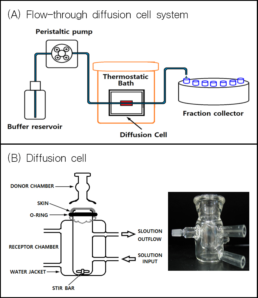
ņĀäņ▓┤ņĀüņØĖ ņŗ£ņŖżĒģ£ņØĆ ņÖäņČ®ņĢĪņØä ņØ╝ņĀĢ ņåŹļÅäļĪ£ diffusion cellļĪ£ Ļ│ĄĻĖēĒĢ┤ ņŻ╝ļŖö ņŚ░ļÅÖĒÄīĒöä(peristaltic pump)ņÖĆ diffusion cellņØś receptor chamberļĪ£ļČĆĒä░ ĒØśļ¤¼ļéśņśżļŖö ņÜ®ņĢĪ(penetrant)ņØä Ļ▓Įņŗ£ļ│äļĪ£ ņłśņ¦æĒĢśļŖö ļČäĒÜŹņłśņ¦æĻĖ░(fraction collector), ĻĘĖļ”¼Ļ│Ā diffusion cellņØä ņØ╝ņĀĢņś©ļÅäļĪ£ ņ£Āņ¦Ćņŗ£ņ╝£ņŻ╝ļŖö ĒĢŁņś©ņĪ░ļĪ£ ļÉśņ¢┤ņ׳ņ£╝ļ®░(Fig. 1), ļČäĒÜŹņłśņ¦æĻĖ░ņŚÉ ņłśņ¦æļÉ£ ņÜ®ņĢĪ ņżæņØś ņĢĮļ¼╝ņØś ļåŹļÅäļŖö HPLCļź╝ ņé¼ņÜ®ĒĢśņŚ¼ ņĀĢļ¤ēļČäņäØņØ┤ Ļ░ĆļŖźĒĢśļŗż. ņŚ¼ĻĖ░ņä£ ņĢĮļ¼╝ņØ┤ Ēö╝ļČĆņĪ░ņ¦üņØä ĒåĄĻ│╝ĒĢśļŖö ņåŹļÅäļŖö ņÖäņČ®ņĢĪņØś ņś©ļÅä, ņ£ĀņåŹ, receptor chamberņØś ļČĆĒö╝ ļ░Å stir barņØś ĻĄÉļ░ś ņåŹļÅäļō▒ņŚÉ ņØśĒĢ┤ ļŗ¼ļØ╝ņ¦ł ņłś ņ׳ļŗż. ļŗ©ņ£ä ņŗ£Ļ░äļ│ä ņĢĮļ¼╝ņØś Ēł¼Ļ│╝ļÉśļŖö ņ¢æņØä Ļ│äņé░ĒĢśļ®┤ ņĢĮļ¼╝ņØś Ēł¼Ļ│╝ ņåŹļÅäļź╝ ĻĄ¼ĒĢĀ ņłś ņ׳ļŗż.
Fig.┬Ā1.
(A): a Flow-through diffusion cell system in which peristaltic pump and fraction collector are connected to diffusion cells, (B) A diffusion cell containing excised skin[9]
2.2 Ēö╝ļČĆļ│┤ĒśĖņĀ£ ņŗ£ĒŚśĒÅēĻ░Ćļź╝ ņ£äĒĢ£ FTD-testņØś ņĪ░Ļ▒┤
Ēö╝ļČĆļ│┤ĒśĖņĀ£ļĪ£ ļÅäĒżļÉ£ Ēö╝ļČĆņĪ░ņ¦ü Ēæ£ļ®┤ņŚÉ ņ£Āņé¼ņ×æņÜ®ņĀ£ļź╝ ļ¢©ņ¢┤ļ£©ļ”¼Ļ│Ā Ēö╝ļČĆĒł¼Ļ│╝ļÅäļź╝ ņĀĢļ¤ēņĀüņ£╝ļĪ£ ĒÅēĻ░ĆĒĢśĻĖ░ņ£äĒĢśņŚ¼ ņŗ£ĒŚśĒÅēĻ░Ć ņĪ░Ļ▒┤ņ£╝ļĪ£ Ļ│ĀļĀżĒĢ┤ņĢ╝ĒĢĀ ņé¼ĒĢŁņØĆ ļŗżņØīĻ│╝ Ļ░Öļŗż.
2.2.1 ņāØņ▓┤ Ēö╝ļČĆņĪ░ņ¦üņØś ņäĀņĀĢ
ļ│Ė ņŗżĒŚśņØä ņ£äĒĢ£ ņŗżĒŚśļÅÖļ¼╝ņØś Ēö╝ļČĆņĪ░ņ¦üņØä ņé¼ņÜ®ĒĢśĻĖ░ ņ£äĒĢśņŚ¼ ņŚ¼ļ¤¼ Ļ░Ćņ¦Ć Ļ│ĀļĀżņÜöņåīĻ░Ć ņ׳ņ£╝ļéś Ļ░Ćņן ņØ┤ņāüņĀüņØĖ ņŗżĒŚśĻ▓░Ļ│╝ļź╝ ņ¢╗ĻĖ░ ņ£äĒĢśņŚ¼ļŖö ņØĖņ▓┤ Ēö╝ļČĆņĪ░ņ¦üĻ│╝ Ļ│äĒåĄļ░£ņāØĒĢÖņĀü(phylogenetically)ņ£╝ļĪ£ Ļ░Ćņן Ļ░ĆĻ╣īņÜ┤ ņŗżĒŚśļÅÖļ¼╝ņØś Ēö╝ļČĆņĪ░ņ¦üņØä ņé¼ņÜ®ĒĢśļŖö Ļ▓āņØ┤ ļ░öļ×īņ¦üĒĢśļŗż. Christopher H. Daltonļō▒ņØĆ[16] ņŗĀĻ▓Įņ×æņÜ®ņĀ£ VXņØś pig skin, human skin, guinea pig skinņŚÉ ļīĆĒĢ£ Ēö╝ļČĆĒł¼Ļ│╝ņ£©ņØä ļ╣äĻĄÉĒĢśĻĖ░ ņ£äĒĢśņŚ¼ FDT-testļź╝ ņŗżņŗ£ĒĢśņśĆļŖöļŹ░, VXņØś 24ņŗ£Ļ░ä ļłäņĀü Ēł¼Ļ│╝ļ¤ēņØ┤ guinea pig skinņØś Ļ▓ĮņÜ░ ņĢĮ 55 mg/cm2, pig skinĻ│╝ human skinņØś Ļ▓ĮņÜ░ļŖö ņĢĮ 10 mg/cm2ļĪ£ ļéśĒāĆļé¼ļŗż. ņ”ē guinea pig skinņØś Ļ▓ĮņÜ░ pig skinĻ│╝ human skin ļ│┤ļŗż ņĢĮ 5ļ░░ ņĀĢļÅä VX Ēł¼Ļ│╝Ļ░Ć ņל ļÉśļ®░, pig skinņØś Ļ▓ĮņÜ░ human skinĻ│╝ Ļ▒░ņØś ņ£Āņé¼ĒĢ£ VX Ēł¼Ļ│╝ļÅäļź╝ ļéśĒāĆļé┤ņŚłļŗż. Simon, G.A.ļō▒ņØĆ[17] pig skinņØ┤ Ļ│äĒåĄļ░£ņāØĒĢÖņĀüņ£╝ļĪ£ļéś ņāØĒÖöĒĢÖņĀüņ£╝ļĪ£ļéś human skinĻ│╝ Ļ░Ćņן Ļ░ĆĻ╣ØļŗżĻ│Ā ļ│┤Ļ│ĀĒĢśņśĆļŖöļŹ░, Christopher H. Daltonļō▒[16]ņØś ņŗżĒŚśĻ▓░Ļ│╝ņÖĆ ņØ╝ņ╣śĒĢ£ļŗżĻ│Ā ļ│╝ ņłś ņ׳ļŗż.
ļ│Ė ņŚ░ĻĄ¼ņŚÉņä£ļŖö 1ņ░©ņĀüņ£╝ļĪ£ hairless mouse skinņØä ņé¼ņÜ®ĒĢ©ņ£╝ļĪ£ņä£ ņŗżĒŚś ļŗ╣ņØ╝ņŚÉ Ēö╝ļČĆļź╝ ņĀüņČ£ĒĢśņŚ¼ Ēö╝ļČĆņĪ░ņ¦üņØś ņł£ĻĖ░ļŖźņØä ņ£Āņ¦ĆĒĢśĻ│Ā Ē¢źĒøä human(cadaver) skinļō▒ņØä ņé¼ņÜ®ĒĢśņŚ¼ ļŹ░ņØ┤Ēä░ļź╝ ĒÖĢņØĖĒĢĀ Ļ│äĒÜŹņØ┤ļŗż. ņäżņ╣śļźś(rodent)ņØś Ēö╝ļČĆņĪ░ņ¦üņØä ņé¼ņÜ®ĒĢśļŖö ļśÉ ĒĢśļéśņØś ņןņĀÉņØĆ ņŗĀĻ▓Į ļ░Å ņłśĒżņ×æņÜ®ņĀ£ņŚÉ ļīĆĒĢ£ in-vivo ņŚ░ĻĄ¼Ļ░Ć Ļ░Ćņן ļ¦ÄņØ┤ ņłśĒ¢ēļÉ£ ņŗżĒŚśļÅÖļ¼╝[18,19]ņØ┤ļ»ĆļĪ£ in-vitro ņŗżĒŚśĻ▓░Ļ│╝ ĒĢ┤ņäØņŚÉ ļÅäņøĆņØ┤ ļÉĀ ņłś ņ׳ļŗż.
2.2.2 ņāØņ▓┤ Ēö╝ļČĆņĪ░ņ¦ü ļīĆņŗĀ ņØĖĻ│Ą membrane ņé¼ņÜ® Ļ░ĆļŖźņä▒
FTD-testļź╝ ņ£äĒĢśņŚ¼ ņāØņ▓┤ Ēö╝ļČĆņĪ░ņ¦üņØä ņé¼ņÜ®ĒĢśļŖö ļīĆņŗĀ ņØĖĻ│ĄņĀüņ£╝ļĪ£ ĒĢ®ņä▒ļÉ£ Ļ│ĀļČäņ×É membraneņØä ņé¼ņÜ®ĒĢ£ ņé¼ļĪĆĻ░Ć ļŗżņłś ļ│┤Ļ│ĀļÉśņŚłļŖöļŹ░[20,21], ņØ┤ļŖö ņŻ╝ļĪ£ ņĢĮļ¼╝ņØś Ēö╝ļČĆ ĒØĪņłśļ│┤ļŗżļŖö ņĢĮļ¼╝ ļ░®ņČ£(drug release) ņŚ░ĻĄ¼ņŚÉ ņŻ╝ļĪ£ ņé¼ņÜ®ļÉ£ļŗż. ņāØņ▓┤ Ēö╝ļČĆņĪ░ņ¦üņØś ņłśĻĖēņØ┤ ņŚ¼ņØśņ╣ś ņĢŖņØä ļĢī ļ╣äņÜ® ņĀłĻ░Éļō▒ņØś ļ¬®ņĀüņ£╝ļĪ£ Ļ│ĀļČäņ×É membraneņØä ņé¼ņÜ®ĒĢśĻ▓ī ļÉśļŖöļŹ░ ņŗżĒŚśņØ┤ Ļ░äĒÄĖĒĢśĻ│Ā ņ×¼ņ¦łņØ┤ ĻĘ£Ļ▓®ĒÖöļÉśņ¢┤ ņ׳ņ£╝ļ»ĆļĪ£ ņŗżĒŚśņØś ņśżņ░©ļ▓öņ£äĻ░Ć ņĀüņØä ņłś ņ׳ļŗżļŖö ņןņĀÉņØ┤ ņ׳ņ£╝ļéś ņ¢┤ļ¢ż membraneņØä ņé¼ņÜ®ĒĢśļŖÉļāÉņŚÉ ļö░ļØ╝ ĻĘĖ ņŗżĒŚśĻ▓░Ļ│╝Ļ░Ć Ēü¼Ļ▓ī ļŗ¼ļØ╝ņ¦ł ņłś ņ׳ļŗż. ĻĘĖļ¤╝ņŚÉļÅä ļČłĻĄ¼ĒĢśĻ│Ā FDAņŚÉņä£ļŖö ņØ╝ļČĆ TDDS ņĀ£ņĀ£ņŚÉ ĒĢ£ņĀĢĒĢśņŚ¼ in-vitro FTD-testļĪ£ ņØĖĻ│Ą membraneņØś ņé¼ņÜ®ņØä ĒŚłĻ░ĆĒĢ£ ļ░ö ņ׳ļŗż[22]. ĻĘĖļ¤¼ļéś ņØ╝ļ░śņĀüņØĖ Ļ▓ĮņÜ░ļŖö ņØĖĻ│Ą membraneņØś ļ¼╝ļ”¼ĒÖöĒĢÖņĀü ĒŖ╣ņä▒ņŚÉ Ļ▓░Ļ│╝Ļ░Ć ņóīņÜ░ļÉśņ¢┤ TDDS ņĀ£ņĀ£ņØś Ēö╝ļČĆ ĒØĪņłśĻ░Ć ņĢäļŗī ņĢĮļ¼╝ ļ░®ņČ£ņØś Ļ▓░Ļ│╝ļĪ£ ļéśĒāĆļé£ļŗżļŖö ņĀ£ĒĢ£ņé¼ĒĢŁņØ┤ ņ׳ļŗż. Simon G.A.ļō▒[21]ņØĆ ņŚ¼ļ¤¼ ņóģļźśņØś ņØĖĻ│Ą membraneĻ│╝ pig ear skinņØä ņé¼ņÜ®ĒĢśņŚ¼ TDDS ņĀ£ņĀ£(Ēī©ņ╣ś)ņŚÉ ļīĆĒĢ£ FDT-testļź╝ ņŗżņŗ£ĒĢśņŚ¼ pig skinĻ│╝ Ļ░Ćņן ĻĘ╝ņĀæĒĢ£ ņØĖĻ│Ą membraneņØä IVIVC(in vivo-in vitro correlation)ņØś Ļ░£ļģÉņØä ņé¼ņÜ®ĒĢśņŚ¼ ņäĀļ│äĒĢśņśĆļŗż. ĻĘĖļ¤¼ļéś J. Milleriouxļō▒[3]ņØĆ ņŗĀĻ▓Įņ×æņÜ®ņĀ£ VXņŚÉ ļīĆĒĢ£ Ēö╝ļČĆļ│┤ĒśĖņĀ£(formulation) 4ņóģņŚÉ ļīĆĒĢ£ Ēö╝ļČĆ ļ│┤ĒśĖņä▒ļŖź ļ╣äĻĄÉļź╝ ņ£äĒĢśņŚ¼ ņŗżļ”¼ņĮś membrane, pig skin, human skinņØä Ļ░üĻ░ü ņé¼ņÜ®ĒĢśņŚ¼ FTD-testļź╝ ņŗżņŗ£ĒĢśņśĆļŖöļŹ░, ņŗżļ”¼ņĮś membraneņØś Ļ▓ĮņÜ░ļŖö Ēö╝ļČĆņĪ░ņ¦üĻ│╝ļŖö ņāüņØ┤ĒĢ£ Ļ▓░Ļ│╝Ļ░Ć ļéśņśżļ»ĆļĪ£ ļ░śļō£ņŗ£ Ēö╝ļČĆņĪ░ņ¦üņØä ņé¼ņÜ®ĒĢśņŚ¼ ĒÖĢņØĖ ņŗżĒŚśņØä ĒĢ┤ņĢ╝ ĒĢ£ļŗżĻ│Ā ļ│┤Ļ│ĀĒĢśņśĆļŗż. ļö░ļØ╝ņä£ Ēö╝ļČĆļ│┤ĒśĖņĀ£ļź╝ ņä▒ļŖźĒÅēĻ░ĆĒĢśĻĖ░ ņ£äĒĢ£ FTD-testņŚÉņä£ļŖö ņŗĀļó░ņä▒ņ׳ļŖö ņŗżĒŚśĻ▓░Ļ│╝ļź╝ ņ¢╗ĻĖ░ņ£äĒĢśņŚ¼ Ļ░ĆĻĖēņĀü ņØĖĻ│Ą membraneņØä ņé¼ņÜ®ĒĢśņ¦Ć ņĢŖļŖö Ļ▓āņØ┤ ļ░öļ×īņ¦üĒĢśļŗżĻ│Ā ĒīÉļŗ©ļÉ£ļŗż.
2.2.3 Ēö╝ļČĆļ│┤ĒśĖņĀ£ ņä▒ļŖźĒÅēĻ░Ćļź╝ ņ£äĒĢ£ FTD-test ņŗżĒŚś ņĪ░Ļ▒┤
ĒÖöĒĢÖņ×æņÜ®ņĀ£ņØś Ēö╝ļČĆ ņ╣©Ēł¼ļź╝ ņ░©ļŗ©ĒĢĀ ņłś ņ׳ļŖö Ēö╝ļČĆļ│┤ĒśĖņĀ£ ņĪ░ņä▒ļ¼╝(formulation)ņŚÉ ļīĆĒĢ£ ņä▒ļŖźņŗżĒŚśņØä ņ£äĒĢśņŚ¼ FTD- testļź╝ ĻĖ░ļ░śņ£╝ļĪ£ ĒĢśļÉś ņČöĻ░ĆņĀüņØĖ ņŗżĒŚśņĪ░Ļ▒┤ņØä ņäżņĀĢĒĢĀ ĒĢäņÜöĻ░Ć ņ׳ļŗż. ņ”ē ņØ╝ļ░śņĀüņØĖ TDDS ņĀ£ņĀ£ņØś ņŗżĒŚśņĪ░Ļ▒┤Ļ│╝ ļŗ¼ļ”¼ ļ│Ė ņŗżĒŚśņŚÉņä£ļŖö TDDS ņĀ£ņĀ£ ļīĆņŗĀ Ēö╝ļČĆļ│┤ĒśĖņĀ£ļź╝ Ēö╝ļČĆņĪ░ņ¦ü ņ£äņŚÉ ĻĘĀņ¦łĒĢśĻ▓ī ļÅäĒżĒĢśĻ│Ā ĻĘĖ ņ£äņŚÉ ņØ╝ņĀĢļ¤ēņØś ņ£Āņé¼ņ×æņÜ®ņĀ£ļź╝ Ļ░ĆĒĢśļŖö Ļ│╝ņĀĢņØ┤ ņČöĻ░ĆļÉ£ļŗż. ņÜ░ņäĀ ņ£Āņé¼ņ×æņÜ®ņĀ£ņØś ņĀüĒĢśļ¤ēņØä Ļ▓░ņĀĢĒĢśĻĖ░ ņ£äĒĢśņŚ¼ ĒāĆ ņŚ░ĻĄ¼ņé¼ļĪĆļź╝ ņé┤ĒÄ┤ļ│┤ļ®┤ ļ»ĖĻĄŁ ECBC(Edgewood Chemical and Biological Center)ņØś JSSED(Joint Service Sensitive Equipment Decontamination)ņŚÉņä£ ņ┤łĻĖ░ņśżņŚ╝ļåŹļÅäļź╝ 10 g/m2(ņĢĮ 1 ╬╝g/cm2)ņ£╝ļĪ£ ņäżņĀĢĒĢśņśĆņ£╝ļ®░[23], ļ»ĖĻĄŁ USAMRICDņŚÉņä£ļŖö aTSP(active Topical Skin Protectant) Ļ░£ļ░£ņØä ņ£äĒĢ┤ ļ│┤ĒśĖņĀ£ in-vitro ņä▒ļŖźņŗ£ĒŚśņŚÉņä£ ņłśĒżņ×æņÜ®ņĀ£ HDļŖö 10 ╬╝Ōäō/cm2, ņŗĀĻ▓Įņ×æņÜ®ņĀ£ GDļŖö 8 ╬╝Ōäō/cm2ļź╝ Ļ░üĻ░ü ņé¼ņÜ®ĒĢśņśĆļŗż[5]. Ēö╝ļČĆĒł¼Ļ│╝ļÅä ņĖĪņĀĢņØä ņ£äĒĢ£ OECD Ļ░ĆņØ┤ļō£ļØ╝ņØĖ[10]ņŚÉņä£ļŖö ņØĖņ▓┤ Ēö╝ļČĆņŚÉ ļīĆĒĢ£ Ēæ£ņżĆ ļģĖņČ£ļ¤ēņØ┤ Ļ│Āņ▓┤ņØś Ļ▓ĮņÜ░ 1-5 mg/cm2, ņĢĪņ▓┤ņØĖ Ļ▓ĮņÜ░ 10 ╬╝Ōäō/cm2ļĪ£ ĻČīņןĒĢśĻ│Ā ņ׳ļŗż. ļö░ļØ╝ņä£ ļ│Ė ņŚ░ĻĄ¼ļź╝ ņ£äĒĢ┤ ņäżĻ│ä ņĀ£ņ×æĒĢ£ diffusion cellņØś Ēö╝ļČĆņĪ░ņ¦ü ļ®┤ņĀü(2 cm2)ņØä Ļ░ÉņĢłĒĢśņŚ¼ FTD- testņŚÉ ņĀüņÜ®ĒĢśĻĖ░ ņ£äĒĢ£ ņ£Āņé¼ņ×æņÜ®ņĀ£ņØś ņ¢æņØĆ 5 - 10 ╬╝Ōäō/cm2ļĪ£ ņäżņĀĢĒĢśņśĆļŗż.
ļśÉĒĢ£ receptor cellņŚÉ ņןņ░®ļÉ£ Ēö╝ļČĆņĪ░ņ¦ü Ēæ£ļ®┤ņŚÉ ļÅäĒżļÉĀ Ēö╝ļČĆļ│┤ĒśĖņĀ£ņØś ļæÉĻ╗śļź╝ Ļ▓░ņĀĢĒĢśĻĖ░ ņ£äĒĢśņŚ¼ ĒāĆ ņŚ░ĻĄ¼ņé¼ļĪĆļź╝ ņé┤ĒÄ┤ļ│┤ļ®┤, ļ»ĖĻĄŁ USAMRICDņØś aTSP ņŚ░ĻĄ¼ņŚÉņä£ļŖö Ēö╝ļČĆļ│┤ĒśĖņĀ£ ļÅäĒżļ¤ēņØ┤ 20 ╬╝g/cm2ļĪ£ņä£ ļæÉĻ╗śļĪ£ ĒÖśņé░ĒĢśļ®┤ ņĢĮ 0.2 mmņŚÉ ĒĢ┤ļŗ╣ļÉ£ļŗż. ļśÉĒĢ£ ļ»ĖĻĄ░ņŚÉņä£ ņÜ┤ņÜ®ļÉ£ Ēö╝ļČĆļ│┤ĒśĖņĀ£ SERPACWAņØś Ļ▓ĮņÜ░ 84 ĻĘĖļש(1ĒÜī ņé¼ņÜ®ļČä Ēżņןļŗ©ņ£ä)ņØä ļģĖņČ£ ņĘ©ņĢĮļČĆņ£ä(ņåÉļ¬®, ļ¬®, ļ░£ļ¬®, Ļ▓©ļō£ļ×æņØ┤, ņé¼ĒāĆĻĄ¼ļŗł, ĒŚłļ”¼ņØś ņ┤Ø ļ®┤ņĀü Ļ│äņé░ņ╣śļŖö ņĢĮ 3,500 cm2)ņŚÉ ļ░öļź┤ļÅäļĪØ ļÉśņ¢┤ ņ׳ļŖöļŹ░ ĒĢ┤ļŗ╣ļÉśļŖö ļæÉĻ╗śļŖö ņĢĮ 0.25 mmļĪ£ Ļ│äņé░ļÉ£ļŗż. ļ│Ė ņŗżĒŚśņŚÉņä£ļŖö ļ╣äĻĄÉņĀü Ļ░ĆĒś╣ņĪ░Ļ▒┤ņØĖ ņŗżņĀ£ ņāüĒÖ®ņŚÉņä£ Ēö╝ļČĆ Ēæ£ļ®┤ņŚÉ ņ×öļźśĒĢĀ ņłś ņ׳ļŖö ļ│┤ĒśĖņĀ£ņØś ņ¢æņØä Ļ░ÉņĢłĒĢśņŚ¼ 0.08 mmĻ░Ć ļÉśļÅäļĪØ ņŗżĒŚśņĪ░Ļ▒┤ņØä ņäżņĀĢĒĢśņśĆļŗż. ĻĘĖ ņÖĖņŚÉ diffusion cellņØś receptor cellņŚÉ Ļ│ĀņĀĢļÉ£ Ēö╝ļČĆņĪ░ņ¦üņØś ņś©ļÅäļź╝ ņŗżņĀ£ ņØĖņ▓┤ņØś Ēö╝ļČĆ Ēæ£ļ®┤ ņś©ļÅäņØĖ 32 ŌäāļĪ£ ļ¦×ņČöĻĖ░ ņ£äĒĢśņŚ¼ diffusion cell ļé┤ņØś ņÜ®ņĢĪ ņś©ļÅäļŖö 37 ŌäāļĪ£ ņ£Āņ¦Ćņŗ£ņ╝░ļŗż[10].
ņŗżĒŚśņ×¼ļŻī ļ░Å ļ░®ļ▓Ģ
3.1 ņŗżĒŚśņ×¼ļŻī
PFPF(Fomblin Y25Ōōć) ļ░Å PTFE(Polymist F5AŌōć, Algoflon L206Ōōć)ļŖö Solvay Inc., glycerinņØĆ C.M. Tech, hairless mouseļŖö ņśżļ”¼ņŚöĒŖĖ ļ░öņØ┤ņśż, perfluoroalkylethyl phosphate(Flexwet PD30Ōōć)ņØĆ Innovative Chemical Technology, polytrimethyl siloxymethacrylate copolymer (FA4103Ōōć)ņØĆ Dow chemical, Nomcort HK-PŌōćļŖö Nisshin Oillio, boron nitrideļŖö Merck, Baycusan 1008ŌōćņØĆ Covestro ņé¼ ņĀ£ĒÆłņØä Ļ░üĻ░ü ņé¼ņÜ®ĒĢśņśĆļŗż. Mg(OH)2, Na2HPO4, methanol(HPLC grade), acetonitrile, sodium alginateļŖö ĻĄŁļé┤ ņĀ£ĒÆł, DMNP, dichlorvosļŖö Aldrich ņĀ£ĒÆłņØä Ļ░üĻ░ü ņé¼ņÜ®ĒĢśņśĆĻ│Ā diffusion cellņØĆ ņŻ╝ļ¼Ė ņĀ£ņ×æĒĢśņŚ¼ ņé¼ņÜ®ĒĢśņśĆļŗż.
3.2 ņŗżĒŚśļ░®ļ▓Ģ
Ēö╝ļČĆļ│┤ĒśĖņĀ£ņØś formulation ņĀ£ņĪ░ļź╝ ļ¬®ņĀüņ£╝ļĪ£ Ļ░ü Ēøäļ│┤ļ¼╝ņ¦łļōżņØś ĻĘĀņ¦łĒÖö Ēś╝ĒĢ® ļ░Å ļČäņé░ņØä ņ£äĒĢśņŚ¼ RobomixerŌōć(high-speed emulsifier/dispenser, T.K PRIMIX, Japan)ļź╝ ņé¼ņÜ®ĒĢśņśĆņ£╝ļ®░, FTD-testļź╝ ĻĖ░ļ░śņ£╝ļĪ£ĒĢ£ Ēö╝ļČĆļ│┤ĒśĖņĀ£ ņä▒ļŖźņŗ£ĒŚśņØä ņ£äĒĢśņŚ¼ ņŗżĒŚśĻ│äĒÜŹņä£ļź╝ ņ×æņä▒ĒĢśĻ│Ā ļÅÖļ¼╝ņŗżĒŚś ņ£żļ”¼ņ£äņøÉĒÜīņØś ņŖ╣ņØĖņØä Ļ▒░ņ│Éņä£ ļŗżņØī ļ░®ļ▓Ģņ£╝ļĪ£ ņŗżņŗ£ĒĢśņśĆļŗż. ņØ┤ņé░ĒÖöĒāäņåīļź╝ ņØ┤ņÜ®ĒĢśņŚ¼ hairless mouse(8ņŻ╝ļĀ╣, ņĢöņ╗Ę)ļź╝ ņĢłļØĮņé¼ņŗ£ĒéżĻ│Ā Ēö╝ļČĆņĪ░ņ¦üņØä ņĀüņČ£ĒĢ£ Ēøä ņ¦Ćļ░®ņ¦łĻ│╝ ĒśłĻ┤ĆņØä ņĀ£Ļ▒░ĒĢśĻ│Ā ņØ╝ņĀĢ ļ®┤ņĀüņ£╝ļĪ£ ņ×ÉļźĖ ļŗżņØī 2 cm2ņØś ņøÉĒśĢ ĻĄ¼ļ®ŹņØ┤ ļܽļ”░ release liner(ļæÉĻ╗ś 0.08 mm)ļź╝ ļīĆĻ│Ā ĻĘĖ ņżæņĢÖņŚÉ ņĖĪņĀĢ ļīĆņāü ņŗ£ļŻī(Ēö╝ļČĆļ│┤ĒśĖņĀ£)ļź╝ 50 - 250 ╬╝Ōäō ļ¢©ņ¢┤ļ£©ļ”░ ļŗżņØī Ēæ£ļ®┤ņŚÉ ņ¢ćĻ▓ī ļ░öļźĖļŗż. ĻĘĖ Ēøä ņŖ¼ļØ╝ņØ┤ļō£ĻĖĆļØ╝ņŖżļź╝ ņØ┤ņÜ®ĒĢśņŚ¼ release liner ļæÉĻ╗śņÖĆ ņØ╝ņ╣śļÉśļÅäļĪØ Ēö╝ļČĆļ│┤ĒśĖņĀ£ļź╝ Ļ╣ÄņĢäļé┤ņ¢┤ Ēæ£ļ®┤ņØä ĒÅēĒāäĒÖöņŗ£ņ╝£ Ēö╝ļČĆļ│┤ĒśĖņĀ£ ņ┤Øļ¤ē(16 mg)ņØ┤ ļÅÖņØ╝ņĪ░Ļ▒┤ņØ┤ ļÉśļÅäļĪØ ĒĢ£ļŗż. ĻĘĖņĀäņŚÉ ļ»Ėļ”¼ multi- channel peristaltic pumpļź╝(sample volume ņĢĮ 3 mŌäō ļÉśĻ▓ī pump ņåŹļÅä ņĪ░ņĀł) ņØ┤ņÜ®ĒĢśņŚ¼ diffusion cellņŚÉ pH 6.0 ņÖäņČ®ņĢĪņØä Ļ│ĄĻĖēĒĢśĻ│Ā circulating water bathļĪ£ diffusion cell ņØś ņś©ļÅäļź╝ 37 ŌäāļĪ£ ņ£Āņ¦Ć ņŗ£ņ╝£ņżĆļŗż. ņĢ×ņä£ ņżĆļ╣äļÉ£ Ēö╝ļČĆļ│┤ĒśĖņĀ£Ļ░Ć ļÅäĒżļÉ£ Ēö╝ļČĆņĪ░ņ¦üņØä diffusion cellņŚÉ ņןņ░®ĒĢ£ Ēøä ņ£Āņé¼ ņ×æņÜ®ņĀ£ļź╝ ņżæņĢÖņŚÉ ļ¢©ņ¢┤ļ£©ļ”¼Ļ│Ā Ēü┤ļשĒöäļĪ£ Ļ│ĀņĀĢņŗ£ņ╝£ņżĆļŗż(Fig. 1B). Diffusion cellņØä ļÆżņ¦æņ¢┤ multi-channel peristaltic pump ņåŹļÅäļź╝ ņĄ£ļīĆļĪ£ ĒĢ£ Ēøä diffusion cell ņĢłņØś ĻĖ░Ēżļź╝ ņĀ£Ļ▒░ĒĢśĻ│Ā multi-channel magnetic stirrer(rpm: 400)ļĪ£ diffusion cell ņĢłņØś ņÜ®ņĢĪņØä ĻĄÉļ░śņŗ£ņ╝£ņżĆļŗż. Ļ▓ĆņĢĪņØĆ 4ņŗ£Ļ░ä Ļ░äĻ▓®ņ£╝ļĪ£ diffusion cellņŚÉņä£ 6 mŌäō ļśÉļŖö 4 mŌäō vialņŚÉ ņŚ░ņåŹņĀüņ£╝ļĪ£ ņ▒äņĘ©ĒĢ£ Ēøä HPLCļź╝ ņé¼ņÜ®ĒĢśņŚ¼ ņĀĢļ¤ēĒĢśļ®░ HPLC ļČäņäØņĪ░Ļ▒┤[24]ņØĆ ļŗżņØīĻ│╝ Ļ░Öļŗż.
ņŗżĒŚśĻ▓░Ļ│╝ ļ░Å ĒĢ┤ņäØ
4.1 ņ£Āņé¼ņ×æņÜ®ņĀ£ DMNP ļ░Å dichlorvosņØś Ēö╝ļČĆ Ēł¼Ļ│╝ļÅä ņ£Āņé¼ņ×æņÜ®ņĀ£ DMNPļŖö GĻ│äņŚ┤ ņŗĀĻ▓Įņ×æņÜ®ņĀ£ņØś ņ£Āņé¼ņ×æņÜ®ņĀ£ļĪ£ ļČäļźśļÉśņ¦Ćļ¦ī ĻĄ¼ņĪ░Ļ░Ć VĻ│äņŚ┤ ņ£Āņé¼ņ×æņÜ®ņĀ£ņØĖ paraoxonĻ│╝ ņ£Āņé¼ĒĢśļ»ĆļĪ£ VĻ│äņŚ┤ ņŗĀĻ▓Įņ×æņÜ®ņĀ£ ņ£Āņé¼ņ×æņÜ®ņĀ£ļĪ£ļÅä ņé¼ņÜ®ļÉ£ļŗż[25,26]. ļśÉĒĢ£ ņŗĀĻ▓Įņ×æņÜ®ņĀ£ ņ£Āņé¼ņ×æņÜ®ņĀ£ dichlorvosņØś Ļ▓ĮņÜ░ GĻ│äņŚ┤ ņŗĀĻ▓Įņ×æņÜ®ņĀ£ņØś ņ£Āņé¼ņ×æņÜ®ņĀ£ļĪ£ņä£ Ļ▓ĮĒö╝ĒØĪņłś(percutaneous absorption) ņŚ░ĻĄ¼ņŚÉ ņ£ĀņÜ®ĒĢśĻ▓ī ņé¼ņÜ®ļÉ£ļŗż[27,28]. ņÜ░ņäĀ Ēö╝ļČĆļ│┤ĒśĖņĀ£ ņŚåņØ┤ Ēö╝ļČĆņĪ░ņ¦ü ņ×Éņ▓┤ņŚÉ ļīĆĒĢ£ ņØ┤ļōż ņ£Āņé¼ņ×æņÜ®ņĀ£ņØś Ēö╝ļČĆĒł¼Ļ│╝ļÅäļź╝ ņĖĪņĀĢĒĢśņśĆļŖöļŹ░ ĻĘĖ Ļ▓░Ļ│╝ļŖö ļŗżņØī Fig. 2ņÖĆ Ļ░Öļŗż. ņ”ē ņ£Āņé¼ņ×æņÜ®ņĀ£ 10 ╬╝Ōäō(ņ”ē DMNP 13.83 mg, dichlorvos 14.20 mg)ļź╝ Ļ░ĆĒĢśņśĆņØä ļĢī 22ņŗ£Ļ░ä ļÅÖņĢłņØś Ēö╝ļČĆ Ēł¼Ļ│╝ļ¤ēņØĆ DMNPĻ░Ć 1.06 mg, dichlorvosĻ░Ć 2.62 mgņ£╝ļĪ£ ļéśĒāĆļé¼ļŗż. ņ”ē dichlorvosļŖö DMNPņŚÉ ļ╣äĒĢ┤ Ēö╝ļČĆĒł¼Ļ│╝ļÅäĻ░Ć 2.5 ļ░░ ņØ┤ņāü Ēü¼ļ®░ ņŗ£Ļ░ä Ļ▓ĮĻ│╝ņŚÉ ļö░ļźĖ Ēö╝ļČĆĒł¼Ļ│╝ profileļÅä dichlorvosņØś Ļ▓ĮņÜ░ ņ▓śņØī 10ņŗ£Ļ░äĻ╣īņ¦ĆļŖö ĻĖēĻ▓®Ē׳ ņ”ØĻ░ĆĒĢśļŗżĻ░Ć ĻĘĖ ņØ┤ĒøäļŖö ņ”ØĻ░ĆĒÅŁņØ┤ ļæöĒÖöļÉśņŚłņ£╝ļ®░ DMNPņØś Ļ▓ĮņÜ░ļŖö ņŗ£Ļ░äņØ┤ ņ¦ĆļéĀņłśļĪØ ņ”ØĻ░ĆĒÅŁņØ┤ ņä£ņä£Ē׳ ņ”ØĻ░ĆĒĢśļŖö ņāüļ░śļÉ£ Ēī©Ēä┤ņØä ļ│┤ņśĆļŗż. ņĢĮļ¼╝ņØś Ēö╝ļČĆĒł¼Ļ│╝ļÅäļź╝ ļ╣äĻĄÉ ņśłņĖĪĒĢśļŖö Ļ▓āņØĆ ņ¢┤ļĀżņÜ┤ ņØ╝ņØ┤ņ¦Ćļ¦ī ļ¼╝ļ”¼ĒÖöĒĢÖņĀü ĒŖ╣ņä▒Ļ│╝ Ēö╝ļČĆņØś ņāØļ”¼ĒĢÖņĀü ĒŖ╣ņä▒ņØä Ļ│ĀļĀżĒĢśņŚ¼ ņ¢┤ļŖÉ ņĀĢļÅäļŖö ņśłņĖĪ Ļ░ĆļŖźĒĢśļŗż. Ēö╝ļČĆĒł¼Ļ│╝ļÅäņŚÉ ņśüĒ¢źņØä ļ»Ėņ╣śļŖö ņŻ╝ņÜöņØĖņ×ÉļŖö ņ£ĀņłśļČäļ░░Ļ│äņłś(µ▓╣µ░┤ÕłåķģŹõ┐éµĢĖ), ļČäņ×Éļ¤ē, ņ£ĄņĀÉ(Ķ׏ķ╗×), ņłś(µ░┤)ņÜ®ĒĢ┤ļÅä, ļČäņ×ÉņØś ĻĄ¼ņĪ░ņĀü ĒŖ╣ņ¦Ģ ļō▒ņØä ļōż ņłś ņ׳ļŖöļŹ░, dichlorvosņÖĆ DMNPļź╝ ņØ┤ļ¤¼ĒĢ£ ņ░©ņøÉņŚÉņä£ ļ╣äĻĄÉĒĢ┤ ļ│┤ļ®┤ ņ£ĀņłśļČäļ░░Ļ│äņłśņØĖ LogP Ļ░ÆņØĆ ĒåĄņāü 2 ~ 3 ņĀĢļÅäĻ░Ć Ēö╝ļČĆĒł¼Ļ│╝ņŚÉ Ļ░Ćņן ņĀüĒĢ®ĒĢśļŗżĻ│Ā ņĢīļĀżņĀĖ ņ׳ņ¢┤ DMNP(1.25)Ļ░Ć dichlorvos(0.71)ļ│┤ļŗż ņÜ░ņäĖĒĢĀ Ļ▓ā ņ£╝ļĪ£ ņśłņĖĪĒĢĀ ņłś ņ׳ļŗż. ĻĘĖļ¤¼ļéś ņ£ĄņĀÉņØĆ dichlorvos(-60 Ōäā)Ļ░Ć DMNP(0 ~ 4 Ōäā)ļ│┤ļŗż ļé«ņĢäņä£ Ēö╝ļČĆĒł¼Ļ│╝ņŚÉ ņ£Āļ”¼ĒĢśļ®░, ļČäņ×Éļ¤ē ņĖĪļ®┤ņŚÉņä£ļÅä ļČäņ×Éļ¤ēņØ┤ ņ×æņØĆ dichlorvosĻ░Ć ņ£Āļ”¼ĒĢśļŗż. DichlorvosņØś ņłśņÜ®ĒĢ┤ļÅäļŖö 8 g/LļĪ£ DMNPņÖĆ ņ£Āņé¼ĒĢ£ ĻĄ¼ņĪ░ļź╝ Ļ░Ćņ¦ä paroxonņØś ņłśņÜ®ĒĢ┤ļÅä(0.5 g/L)ļź╝ Ļ│ĀļĀż ņŗ£ dichlorvosņØś ņłśņÜ®ĒĢ┤ļÅäĻ░Ć ļŹö ļåÆņØä Ļ▓āņ£╝ļĪ£ ņśłņĖĪļÉśņ¢┤ Ēö╝ļČĆĒł¼Ļ│╝ņŚÉ ļ│┤ļŗż ņ£Āļ”¼ĒĢśļŗż. ļ│äļÅäļĪ£ ļ│Ė ņŗżĒŚśņĪ░Ļ▒┤ņŚÉņä£ DMNPņØś Ļ░ĆņłśļČäĒĢ┤ĒÜ©Ļ│╝ļź╝ ĒÖĢņØĖĒĢśņśĆļŖöļŹ░ 37 ŌäāņŚÉņä£ 24ņŗ£Ļ░ä ļÅÖņĢł 0.04 %Ļ░Ć ļČäĒĢ┤ļÉśņ¢┤ Ļ░ĆņłśļČäĒĢ┤ĒÜ©Ļ│╝ļŖö ņŗżĒŚśņŚÉ ņśüĒ¢źņØ┤ Ļ▒░ņØś ņŚåļŖö Ļ▓āņ£╝ļĪ£ ĒÖĢņØĖļÉśņŚłļŗż.

4.2 ļ»ĖĻĄŁ Ēö╝ļČĆļ│┤ĒśĖņĀ£ SERPACWAņØś ņŻ╝ņÜö ņä▒ļČäņØś ĒÜ©ļŖź ļČäņäØ
ļ»ĖĻĄŁ USAMRICDņŚÉņä£ ņŚ░ĻĄ¼Ļ░£ļ░£ļÉ£ Ēö╝ļČĆļ│┤ĒśĖņĀ£ SERPACWAļŖö MOPP ņāüĒÖ®ņŚÉņä£ ļģĖņČ£ņŚÉ ņĘ©ņĢĮĒĢ£ Ēö╝ļČĆ ļČĆņ£äņŚÉ ļ»Ėļ”¼ ļ░£ļØ╝ņä£ ĒÖöĒĢÖņ×æņÜ®ņĀ£ļĪ£ļČĆĒä░ Ēö╝ļČĆļź╝ ļ│┤ĒśĖĒĢśļŖö ņŚ░Ļ│ĀņĀ£ļĪ£ņä£ ņŻ╝ņä▒ļČäņØĆ PFPE(perfluorinated polyether, Fomblin Y25Ōōć, ņśżņØ╝ ņä▒ļČä)ņÖĆ PTFE(polytetrafluoroethylene, Polymist F5AŌōć, ļ»ĖņäĖļČäļ¦É)ņØś 1:1 ļ░░ĒĢ®(w/w)ņ£╝ļĪ£ ņØ┤ļŻ©ņ¢┤ņĀĖ ņ׳ļŗż.
ļö░ļØ╝ņä£ PTFE ĒĢ©ļ¤ēņŚÉ ļö░ļźĖ Ēö╝ļČĆļ│┤ĒśĖĒÜ©Ļ│╝ļź╝ ņŗ£ĒŚśĒĢ┤ļ│┤Ļ│Āņ×É PFPE 1.0 ļīĆļ╣ä PTFE 0.1, 0.3, 0.5, 0.7, 1.0ņØś 5Ļ░Ćņ¦Ć Ēś╝ĒĢ®ļ╣äņ£©ļĪ£ ņĪ░ņä▒ļ¼╝ņØä ņĀ£ņĪ░ĒĢśņŚ¼ ņÜ░ņäĀ M9 ĒāÉņ¦Ćņ¦ĆļĪ£ ņŗżĒŚśĒĢśņŚ¼ ļ│┤ņĢśļŖöļŹ░ PTFE ļåŹļÅäņŚÉ ļ╣äļĪĆĒĢśņŚ¼ ļ│┤ĒśĖņä▒ļŖź ņ”ØĻ░ĆĻ░Ć Ļ┤Ćņ░░ļÉśņŚłļŖöļŹ░ ņØ┤ļź╝ ĒÖĢņØĖĒĢśĻ│Āņ×É ļÅÖņØ╝ ņĪ░ņä▒ļ¼╝ņŚÉ ļīĆĒĢśņŚ¼ FTD-testļź╝ ņŗżņŗ£ĒĢśņśĆĻ│Ā ĻĘĖ Ļ▓░Ļ│╝ļŖö Fig. 3Ļ│╝ Ļ░Öļŗż. Fig. 3ņŚÉņä£ PTFEņØś ĒĢ©ļ¤ēņØ┤ ņ”ØĻ░ĆļÉĀņłśļĪØ ņØ┤ņÖĆ ļ╣äļĪĆĒĢśņŚ¼ ņ£Āņé¼ņ×æņÜ®ņĀ£ DMNPņŚÉ ļīĆĒĢ£ ļ│┤ĒśĖņä▒ļŖźņØ┤ ņ”ØĻ░ĆĒĢ©ņØä Ļ┤Ćņ░░ĒĢśņśĆĻ│Ā, ļÅÖņØ╝ĒĢ£ ļ░®ļ▓ĢĻ│╝ ņĪ░Ļ▒┤ņ£╝ļĪ£ 5Ļ░Ćņ¦Ć ņĪ░ņä▒ļ¼╝ņŚÉ ļīĆĒĢśņŚ¼ ņ£Āņé¼ņ×æņÜ®ņĀ£ dichlorvosļź╝ ņé¼ņÜ®ĒĢśņŚ¼ ņŗżĒŚśĒĢśņśĆļŖöļŹ░ ĻĘĖ Ļ▓░Ļ│╝ļŖö ļŗżņØī Fig. 4ņÖĆ Ļ░Öļŗż. Fig. 4ņŚÉņä£ dichlorvos ņØś Ļ▓ĮņÜ░ ļ│ĄĒĢ® ņĪ░ņä▒ļ¼╝ņØś ļ│┤ĒśĖņä▒ļŖźļÅä DMNPņŚÉ ļ╣äĒĢ┤ Ēü¼Ļ▓ī Ļ░ÉņåīĒĢśļŖö Ļ▓āņ£╝ļĪ£ ļéśĒāĆļé¼ļŗż. ņØ┤ļŖö ņ£Āņé¼ņ×æņÜ®ņĀ£ dichlorvosņØś Ēö╝ļČĆņ╣©Ēł¼ ņÜ®ņØ┤ņä▒ ļĢīļ¼ĖņŚÉ Ēö╝ļČĆļ│┤ĒśĖņĀ£ņØś ļ│┤ĒśĖņä▒ļŖźņØ┤ ļŗżņåī ņĀĆĒĢśļÉĀ ņłś ņ׳ļŖö ņĪ░Ļ▒┤ņØ┤ļ»ĆļĪ£ dichlorvos ļŖö DMNPņŚÉ ļ╣äĒĢśņŚ¼ Ēö╝ļČĆļ│┤ĒśĖņĀ£ņØś ļ│┤ĒśĖņä▒ļŖźĒÅēĻ░ĆņŚÉ ĒÜ©Ļ│╝ņĀüņ£╝ļĪ£ ņé¼ņÜ®Ļ░ĆļŖźĒĢ£ ņ£Āņé¼ņ×æņÜ®ņĀ£ļĪ£ ĒīÉļŗ©ļÉ£ļŗż.

4.3 ļ»ĖĻĄŁ Ēö╝ļČĆļ│┤ĒśĖņĀ£ SERPACWAņÖĆ ņØ┤ņŖżļØ╝ņŚś Ēö╝ļČĆļ│┤ĒśĖņĀ£ IB-1ņØś ņŻ╝ņÜö ņä▒ļČä ļ╣äĻĄÉņŗżĒŚś
ļ│┤ĒśĖņĀ£ IB-1ņØś ņŻ╝ņÜö ņä▒ļČä ļ╣äĻĄÉņŗżĒŚś Ēö╝ļČĆļ│┤ĒśĖņĀ£ IB-1(ņāüĒÆłļ¬ģ DermostyxŌōć)ļŖö IIBR(Israel Institute of Biological Research)ņŚÉņä£ Ļ░£ļ░£ļÉśņ¢┤ Ēśäņ×¼ ņØ┤ņŖżļØ╝ņŚśņŚÉņä£ ņÜ┤ņÜ® ņżæņØĖ ĻĄ░ņÜ® Ēö╝ļČĆļ│┤ĒśĖņĀ£ļĪ£ņä£ ņŻ╝ņä▒ļČäņØĆ glycerinņØĖ Ļ▓āņ£╝ļĪ£ ņĢīļĀżņĪīņ£╝ļ®░, IB-1ņØś ņÜ░ņłśĒĢ£ Ēö╝ļČĆļ│┤ĒśĖņä▒ļŖźņŚÉ ļīĆĒĢ┤ ļŗżņłśņØś ņŚ░ĻĄ¼Ļ▓░Ļ│╝[29-31]Ļ░Ć ļ│┤Ļ│ĀļÉ£ ļ░ö ņ׳ļŗż. ļö░ļØ╝ņä£ glycerinņØä ņŻ╝ņä▒ļČäņ£╝ļĪ£ ĒĢśļ®┤ņä£ ņĀÉļÅäļŖö ļŹöņÜ▒ ņ”ØĻ░Ćņŗ£ĒéżĻ│Ā ņāüļČäļ”¼ ļ░Å ņ╣©ņĀäĒśäņāüņØ┤ ņŚåļŖö ļ░▒ņāēĒü¼ļ”╝ņØä ņĀ£ņĪ░ĒĢśņŚ¼ ļ»ĖĻĄŁ SERPACWAņØś ņŻ╝ņÜöņä▒ļČäĻ│╝ ļ│┤ĒśĖņä▒ļŖźņØä ļ╣äĻĄÉĒĢśņŚ¼ ļ│┤ņĢśļŖöļŹ░ ĻĘĖ Ļ▓░Ļ│╝ļŖö ļŗżņØī Table 1Ļ│╝ Ļ░Öļŗż.
Table┬Ā1.
Percent of protection provided by TSP formulations with various compositions against percutaneous absorption of DMNP. (A22hr : cumulative total amount(╬╝g) of penetrant for 22 hours, D initial : Initial dose (╬╝g) of DMNP applied on skin)
| Formulations Compositions | TSP | TSP-1 | IB-1 | PSB | GBD-2 |
|---|---|---|---|---|---|
| Fomblin Y25 | 50 | 50 | - | - | - |
| Polymist F5A | 50 | - | - | - | - |
| Algoflon L206 | - | 50 | - | - | - |
| Glycerin | - | - | 90 | 17.8 | 74.8 |
| Mica | - | - | 10 | - | - |
| Sodium alginate | - | - | - | 4.4 | - |
| FA4103 | - | - | - | - | 9.7 |
| Noncomport HK-P | - | - | - | - | 5.8 |
| Boron nitride | - | - | - | - | 9.7 |
| Water | - | - | - | 77.8 | - |
| Percent of protectionŌłŚ | 99.9 | 99.9 | 98.4 | 98.8 | 98.0 |
Table 1ņŚÉņä£ ļ»ĖĻĄŁ SERPACWAņØś ņŻ╝ņÜö ņä▒ļČäņØĖ ļČłņåīĻ│ä ĒÖöĒĢ®ļ¼╝ņØä ņŻ╝ņä▒ļČäņ£╝ļĪ£ĒĢśļŖö ņ▓śļ░®(TSP, TSP-1)ņØ┤ IB-1ņØś ņŻ╝ņä▒ļČäņØĖ glycerinņØä ņŻ╝ņä▒ļČäņ£╝ļĪ£ĒĢśļŖö ņ▓śļ░®(IB-1, PSB, GBD-2)ļ│┤ļŗż ņ£Āņé¼ņ×æņÜ®ņĀ£ DMNPņŚÉ ļīĆĒĢśņŚ¼ ņÜ░ņłśĒĢ£ Ēö╝ļČĆ ļ│┤ĒśĖĒÜ©Ļ│╝ļź╝ ļéśĒāĆļé┤ņŚłļŗż. TSPņÖĆ TSP-1ņ£╝ļĪ£ ļÅäĒżļÉ£ ņŗ£ĒŚśĻĄ░ņØś Ļ▓ĮņÜ░ glycerinņØä ĒżĒĢ©ĒĢśļŖö ņĪ░ņä▒ļ¼╝Ļ│╝ļŖö ļŗ¼ļ”¼ ņ£Āņé¼ņ×æņÜ®ņĀ£ DMNPĻ░Ć Ēæ£ļ®┤ņŚÉ ĒŹ╝ņ¦Ćņ¦ĆņĢŖĻ│Ā ļ░®ņÜĖ ņāüĒā£ļĪ£ ļ¦║Ē׳ļŖö ĒśäņāüņØ┤ Ļ┤Ćņ░░ļÉśņŚłņ£╝ļ®░ ņØ┤ņÖĆĻ░ÖņØĆ ĒśäņāüņØ┤ ļ│┤ĒśĖņä▒ļŖźņØä ļåÆņØ╝ ņłś ņ׳ļŖö ĒĢ£Ļ░Ćņ¦Ć ņøÉņØĖņ£╝ļĪ£ ņé¼ļŻīļÉ£ļŗż.
Ļ│Ā ņ░░
ļ»ĖĻĄŁņØś Ēö╝ļČĆļ│┤ĒśĖņĀ£ņØĖ SERPACWAņØś ņŻ╝ņä▒ļČäņØĖ PFPE ņÖĆ PTFEļŖö ņŻ╝ļĪ£ ĒāäņåīņÖĆ ļČłņåīļĪ£ ņØ┤ļŻ©ņ¢┤ņ¦ä Ļ│ĀļČäņ×É ĒÖöĒĢ®ļ¼╝ļĪ£ņä£ ņśżļל ņĀäļČĆĒä░ ĒÖöņןĒÆł ļ░Å ņ£żĒÖ£ņ£ĀņØś ņøÉļŻīļĪ£ ņé¼ņÜ®ļÉśņŚłļŗż. PFPEļŖö ĒÖöĒĢÖņĀüņ£╝ļĪ£ ļ¦żņÜ░ ņĢłņĀĢĒĢśņŚ¼ ņØĖņ▓┤ņŚÉ ļÅģņä▒ņØ┤ Ļ▒░ņØś ņŚåņ£╝ļ®░ ņ¢┤ļ¢ĀĒĢ£ ņ£ĀĻĖ░ņÜ®ļ¦żņŚÉļÅä ļģ╣ņ¦ĆņĢŖļŖö ĒŖ╣ņä▒ņØä Ļ░Ćņ¦ĆĻ│Ā ņ׳ļŗż. ĒŖ╣ņä▒ņØä ņÜöņĢĮĒĢ┤ļ│┤ļ®┤ ņåīņłśņä▒ņØ┤ņ¦Ćļ¦ī ņ¦Ćļ░®ņĖĄĻ│╝ļÅä ņä×ņØ┤ņ¦Ć ņĢŖļŖö, ņ”ē ļ¼╝Ļ│╝ ĻĖ░ļ”äņŚÉ ļ¬©ļæÉ ņä×ņØ┤ņ¦Ć ņĢŖņ£╝ļ®░ PFPE Ļ░äņŚÉļÅä ņä£ļĪ£ ļ░śļ░£ĒĢśļŖö ņä▒ņ¦łņØ┤ ņ׳ļŗż. ņØ┤ņÖĆ Ļ░ÖņØĆ ĒŖ╣ņä▒ņ£╝ļĪ£ ļ╣äĻĄÉņĀü ņåīņłśņä▒ņŚÉ Ļ░ĆĻ╣īņÜ┤ ĒÖöĒĢÖņ×æņÜ®ņĀ£ņŚÉ ļīĆĒĢ£ Ēö╝ļČĆ ļ│┤ĒśĖĒÜ©Ļ│╝Ļ░Ć glycerin ļ│┤ļŗż ņÜ░ņłśĒĢ£ Ļ▓āņ£╝ļĪ£ ĒĢ┤ņäØļÉ£ļŗż. ĻĘĖļ¤¼ļéś ņ£Āņé¼ņ×æņÜ®ņĀ£Ļ░äņØś Ēö╝ļČĆĒł¼Ļ│╝ļÅäĻ░Ć ņāüņØ┤ĒĢśĻ│Ā ņ£Āņé¼ņ×æņÜ®ņĀ£-Ēö╝ļČĆļ│┤ĒśĖņĀ£Ļ░äņØś ļ¼╝ļ”¼ĒÖöĒĢÖņĀü ņāüĒśĖņ×æņÜ®ņØ┤ ļŗżļź╝ ņłś ņ׳ĻĖ░ ļĢīļ¼ĖņŚÉ Ē¢źĒøä ļŗżņ¢æĒĢ£ ņóģļźśņØś ņ£Āņé¼ņ×æņÜ®ņĀ£ ļ░Å ņŗżņ×æņÜ®ņĀ£ņŚÉ ļīĆĒĢśņŚ¼ FTD-testļź╝ ņŗżņŗ£ĒĢĀ ĒĢäņÜöņä▒ņØ┤ ņ׳ņØä Ļ▓āņ£╝ļĪ£ ĒīÉļŗ©ļÉ£ļŗż.
Ļ▓░ ļĪĀ
TDDS ņĀ£ņĀ£ņØś ņĢĮļ¼╝ ļ░®ņČ£ ļ░Å ņĢĮļ¼╝ļÅÖļĀźĒĢÖņĀü ņŚ░ĻĄ¼ņŚÉ ņé¼ņÜ®ļÉśļŖö FTD-testļź╝ ĻĖ░ļ░śņ£╝ļĪ£ ĒÖöĒĢÖņ×æņÜ®ņĀ£ņŚÉ ļīĆĒĢ£ Ēö╝ļČĆļ│┤ĒśĖņĀ£ļź╝ ņä▒ļŖźĒÅēĻ░ĆĒĢĀ ņłś ņ׳ļŖö ņŗ£ĒŚśĒÅēĻ░Ćļ▓ĢņØä ĻĄŁļé┤ ņĄ£ņ┤łļĪ£ ĒÖĢļ”ĮĒĢśņśĆļŗż. ĒÖĢļ”ĮļÉ£ ņŗ£ĒŚśĒÅēĻ░Ć ļ░®ļ▓ĢņŚÉ ņØśĒĢ┤ ļ»ĖĻĄŁ ļ░Å ņØ┤ņŖżļØ╝ņŚśņØś Ēö╝ļČĆļ│┤ĒśĖņĀ£ņŚÉ ņé¼ņÜ®ļÉ£ ņŻ╝ņÜö ņä▒ļČäļōżņØä ņ£äņŻ╝ļĪ£ ļ¬ćĻ░Ćņ¦Ć ļ│ĄĒĢ®ņĪ░ņä▒ļ¼╝(formulation)ņØä ņĀ£ņĪ░ĒĢśņśĆĻ│Ā ņØ┤ļōżņŚÉ ļīĆĒĢ£ Ēö╝ļČĆļ│┤ĒśĖņä▒ļŖźņØä ļ╣äĻĄÉ ĒÅēĻ░ĆĒĢśņŚ¼ ņ£ĀņØśņä▒ ņ׳ļŖö Ļ▓░Ļ│╝ļź╝ ĒÖĢņØĖĒĢśņśĆļŗż. ļ│Ė ņŚ░ĻĄ¼ņŚÉ ņé¼ņÜ®ļÉ£ ņ£Āņé¼ņ×æņÜ®ņĀ£ņØś Ļ▓ĮņÜ░ ņłśņÜ®ņĢĪ ņāüņŚÉņä£ Ļ░ĆņłśļČäĒĢ┤ņ£©ņØ┤ ļ¦żņÜ░ ļé«ņĢä(24ņŗ£Ļ░äņŚÉ 0.04 % ņłśņżĆ) ņŗżĒŚśĻ▓░Ļ│╝ņŚÉ ņśüĒ¢źņØä ņŻ╝ņ¦Ć ņĢŖņĢśņ£╝ļ®░, ņŗĀĻ▓Įņ×æņÜ®ņĀ£ VXņØś Ļ▓ĮņÜ░ļÅä ņłśņÜ®ņĢĪņŚÉņä£ ļ╣äĻĄÉņĀü ņĢłņĀĢĒĢśļ»ĆļĪ£ ņ£Āņé¼ĒĢ£ ņĪ░Ļ▒┤ņŚÉņä£ ņŗżĒŚśņØ┤ Ļ░ĆļŖźĒĢśļŗż[3,16]. ĻĘĖļ¤¼ļéś ņŗĀĻ▓Įņ×æņÜ®ņĀ£ GBņØś Ļ▓ĮņÜ░ļŖö ļČäĒĢ┤ņé░ļ¼╝ ņāØņä▒ņØä Ļ│ĀļĀżĒĢ£ ņŗżĒŚśņĪ░Ļ▒┤ņØś ņäżņĀĢņØ┤ ĒĢäņÜöĒĢĀ Ļ▓āņ£╝ļĪ£ ĒīÉļŗ©ļÉ£ļŗż. ļö░ļØ╝ņä£ ļ│Ė ņŚ░ĻĄ¼ņŚÉņä£ ĒÖĢļ”ĮļÉ£ ņŗ£ĒŚśĒÅēĻ░ĆņĪ░Ļ▒┤ņØĆ Ē¢źĒøä ĒÖöĒĢÖņ×æņÜ®ņĀ£ņŚÉ ļīĆĒĢ£ Ēö╝ļČĆļ│┤ĒśĖņĀ£ņØś ņŗ£ĒŚśĒÅēĻ░ĆņŚÉ ņĀüņÜ® Ļ░ĆļŖźĒĢ£ ĒĢśļéśņØś Ēæ£ņżĆņŗ£ĒŚśņĀłņ░©ņä£(SOP; Standard Operational Procedure)ļĪ£ ņé¼ņÜ® Ļ░ĆļŖźĒĢĀ Ļ▓āņ£╝ļĪ£ ĒīÉļŗ©ļÉ£ļŗż.


